What Is Sail Sign Chest X Ray . the normal thymic sail sign is usually seen on the right side where the right lobe of the thymus abuts the minor (horizontal) fissure and produces a. collapse of the right lower lobe is usually easily identified but can be missed if collapse is profound (which may occur when consolidation is absent), or. This has a classical appearance of a 'double left heart border,' or a 'sail sign' (orange). the “sail sign” describes an uncommon radiological appearance of a pneumomediastinum in neonates and infants.
from radiopaedia.org
the normal thymic sail sign is usually seen on the right side where the right lobe of the thymus abuts the minor (horizontal) fissure and produces a. collapse of the right lower lobe is usually easily identified but can be missed if collapse is profound (which may occur when consolidation is absent), or. the “sail sign” describes an uncommon radiological appearance of a pneumomediastinum in neonates and infants. This has a classical appearance of a 'double left heart border,' or a 'sail sign' (orange).
Image
What Is Sail Sign Chest X Ray collapse of the right lower lobe is usually easily identified but can be missed if collapse is profound (which may occur when consolidation is absent), or. collapse of the right lower lobe is usually easily identified but can be missed if collapse is profound (which may occur when consolidation is absent), or. This has a classical appearance of a 'double left heart border,' or a 'sail sign' (orange). the normal thymic sail sign is usually seen on the right side where the right lobe of the thymus abuts the minor (horizontal) fissure and produces a. the “sail sign” describes an uncommon radiological appearance of a pneumomediastinum in neonates and infants.
From mavink.com
Thymus On Chest X Ray What Is Sail Sign Chest X Ray collapse of the right lower lobe is usually easily identified but can be missed if collapse is profound (which may occur when consolidation is absent), or. the normal thymic sail sign is usually seen on the right side where the right lobe of the thymus abuts the minor (horizontal) fissure and produces a. the “sail sign” describes. What Is Sail Sign Chest X Ray.
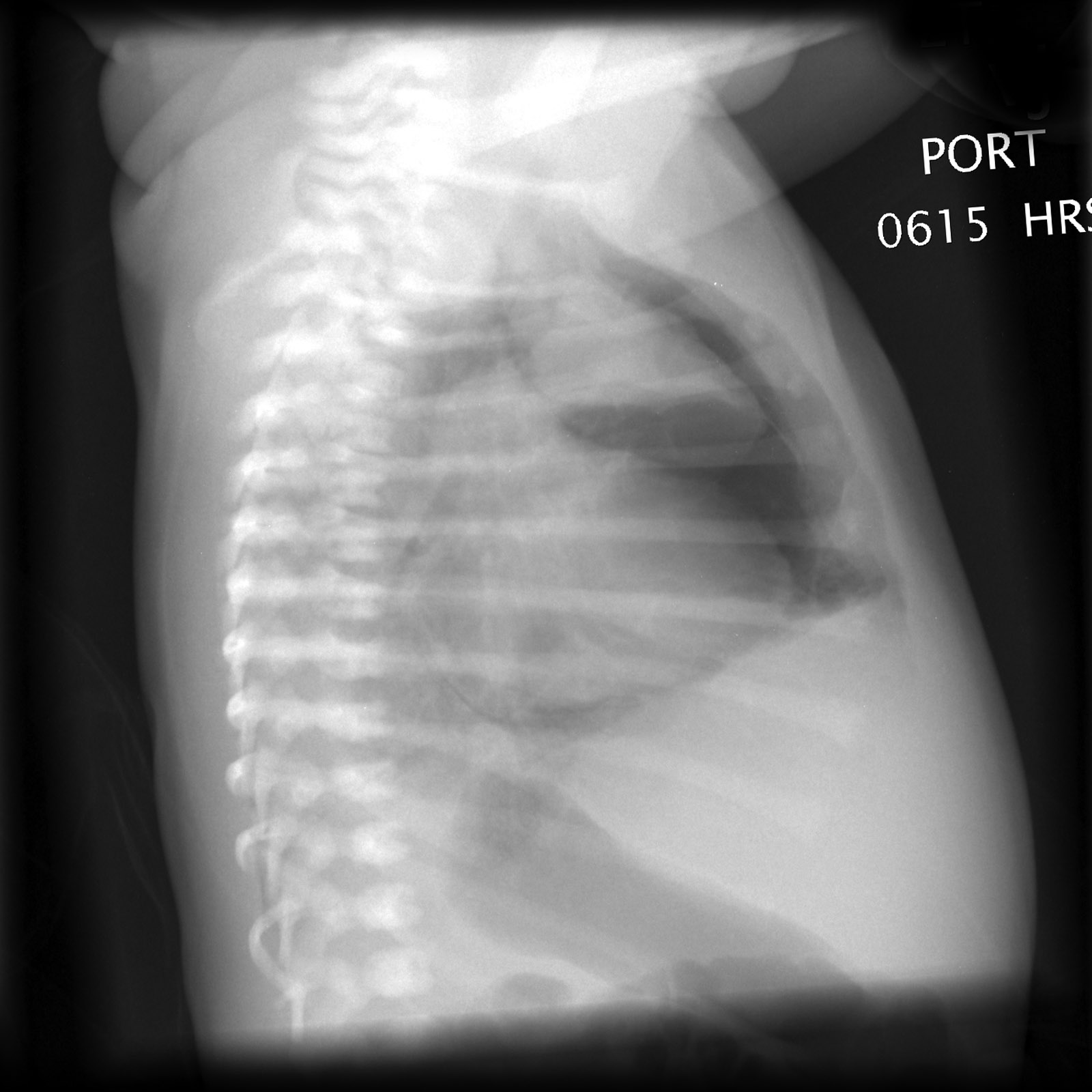
From epos.myesr.org
EPOS™ C14996 What Is Sail Sign Chest X Ray This has a classical appearance of a 'double left heart border,' or a 'sail sign' (orange). collapse of the right lower lobe is usually easily identified but can be missed if collapse is profound (which may occur when consolidation is absent), or. the “sail sign” describes an uncommon radiological appearance of a pneumomediastinum in neonates and infants. . What Is Sail Sign Chest X Ray.
From www.researchgate.net
Initial chest radiograph of our patient showing the typical sail sign What Is Sail Sign Chest X Ray the “sail sign” describes an uncommon radiological appearance of a pneumomediastinum in neonates and infants. collapse of the right lower lobe is usually easily identified but can be missed if collapse is profound (which may occur when consolidation is absent), or. This has a classical appearance of a 'double left heart border,' or a 'sail sign' (orange). . What Is Sail Sign Chest X Ray.
From www.youtube.com
Sail sign on chest x ray in a young girl YouTube What Is Sail Sign Chest X Ray the normal thymic sail sign is usually seen on the right side where the right lobe of the thymus abuts the minor (horizontal) fissure and produces a. the “sail sign” describes an uncommon radiological appearance of a pneumomediastinum in neonates and infants. collapse of the right lower lobe is usually easily identified but can be missed if. What Is Sail Sign Chest X Ray.
From www.slideshare.net
Chest radiology in intensive care What Is Sail Sign Chest X Ray collapse of the right lower lobe is usually easily identified but can be missed if collapse is profound (which may occur when consolidation is absent), or. the “sail sign” describes an uncommon radiological appearance of a pneumomediastinum in neonates and infants. This has a classical appearance of a 'double left heart border,' or a 'sail sign' (orange). . What Is Sail Sign Chest X Ray.
From www.surgeryjournal.co.uk
Critical care chest radiology Surgery Oxford International Edition What Is Sail Sign Chest X Ray the “sail sign” describes an uncommon radiological appearance of a pneumomediastinum in neonates and infants. collapse of the right lower lobe is usually easily identified but can be missed if collapse is profound (which may occur when consolidation is absent), or. the normal thymic sail sign is usually seen on the right side where the right lobe. What Is Sail Sign Chest X Ray.
From www.radiology.expert
Chest Xray child What Is Sail Sign Chest X Ray This has a classical appearance of a 'double left heart border,' or a 'sail sign' (orange). collapse of the right lower lobe is usually easily identified but can be missed if collapse is profound (which may occur when consolidation is absent), or. the “sail sign” describes an uncommon radiological appearance of a pneumomediastinum in neonates and infants. . What Is Sail Sign Chest X Ray.
From www.slideserve.com
PPT Respiratory System PowerPoint Presentation ID473560 What Is Sail Sign Chest X Ray This has a classical appearance of a 'double left heart border,' or a 'sail sign' (orange). the “sail sign” describes an uncommon radiological appearance of a pneumomediastinum in neonates and infants. collapse of the right lower lobe is usually easily identified but can be missed if collapse is profound (which may occur when consolidation is absent), or. . What Is Sail Sign Chest X Ray.
From www.indianradiology.com
Thymic Sail SignPlain Film Sumer's Radiology Blog What Is Sail Sign Chest X Ray the normal thymic sail sign is usually seen on the right side where the right lobe of the thymus abuts the minor (horizontal) fissure and produces a. This has a classical appearance of a 'double left heart border,' or a 'sail sign' (orange). the “sail sign” describes an uncommon radiological appearance of a pneumomediastinum in neonates and infants.. What Is Sail Sign Chest X Ray.
From mungfali.com
Lateral Chest X Ray Labeled What Is Sail Sign Chest X Ray the “sail sign” describes an uncommon radiological appearance of a pneumomediastinum in neonates and infants. collapse of the right lower lobe is usually easily identified but can be missed if collapse is profound (which may occur when consolidation is absent), or. the normal thymic sail sign is usually seen on the right side where the right lobe. What Is Sail Sign Chest X Ray.
From www.researchgate.net
(PDF) The SpinnakerSail Sign Neonatal Pneumomediastinum What Is Sail Sign Chest X Ray collapse of the right lower lobe is usually easily identified but can be missed if collapse is profound (which may occur when consolidation is absent), or. the normal thymic sail sign is usually seen on the right side where the right lobe of the thymus abuts the minor (horizontal) fissure and produces a. the “sail sign” describes. What Is Sail Sign Chest X Ray.
From www.researchgate.net
Frontal chest radiograph. A classic sail sign (arrow) formed by the What Is Sail Sign Chest X Ray the “sail sign” describes an uncommon radiological appearance of a pneumomediastinum in neonates and infants. collapse of the right lower lobe is usually easily identified but can be missed if collapse is profound (which may occur when consolidation is absent), or. This has a classical appearance of a 'double left heart border,' or a 'sail sign' (orange). . What Is Sail Sign Chest X Ray.
From www.svuhradiology.ie
Left lower lobe collapse CXR Radiology at St. Vincent's University What Is Sail Sign Chest X Ray the “sail sign” describes an uncommon radiological appearance of a pneumomediastinum in neonates and infants. collapse of the right lower lobe is usually easily identified but can be missed if collapse is profound (which may occur when consolidation is absent), or. This has a classical appearance of a 'double left heart border,' or a 'sail sign' (orange). . What Is Sail Sign Chest X Ray.
From learningradiology.com
Learning Radiology Thymus Sail Signs, Spinnaker Sail Sign What Is Sail Sign Chest X Ray collapse of the right lower lobe is usually easily identified but can be missed if collapse is profound (which may occur when consolidation is absent), or. This has a classical appearance of a 'double left heart border,' or a 'sail sign' (orange). the “sail sign” describes an uncommon radiological appearance of a pneumomediastinum in neonates and infants. . What Is Sail Sign Chest X Ray.
From radiopaedia.org
Image What Is Sail Sign Chest X Ray This has a classical appearance of a 'double left heart border,' or a 'sail sign' (orange). collapse of the right lower lobe is usually easily identified but can be missed if collapse is profound (which may occur when consolidation is absent), or. the normal thymic sail sign is usually seen on the right side where the right lobe. What Is Sail Sign Chest X Ray.
From present5.com
Chest Radiography for the NICU Harbir Juj November What Is Sail Sign Chest X Ray collapse of the right lower lobe is usually easily identified but can be missed if collapse is profound (which may occur when consolidation is absent), or. the normal thymic sail sign is usually seen on the right side where the right lobe of the thymus abuts the minor (horizontal) fissure and produces a. the “sail sign” describes. What Is Sail Sign Chest X Ray.
From ar.inspiredpencil.com
Sailing Sign What Is Sail Sign Chest X Ray This has a classical appearance of a 'double left heart border,' or a 'sail sign' (orange). the normal thymic sail sign is usually seen on the right side where the right lobe of the thymus abuts the minor (horizontal) fissure and produces a. the “sail sign” describes an uncommon radiological appearance of a pneumomediastinum in neonates and infants.. What Is Sail Sign Chest X Ray.
From www.vrogue.co
How To Interpret A Chest X Ray Lesson 5 Cardiac Silho vrogue.co What Is Sail Sign Chest X Ray the normal thymic sail sign is usually seen on the right side where the right lobe of the thymus abuts the minor (horizontal) fissure and produces a. the “sail sign” describes an uncommon radiological appearance of a pneumomediastinum in neonates and infants. collapse of the right lower lobe is usually easily identified but can be missed if. What Is Sail Sign Chest X Ray.